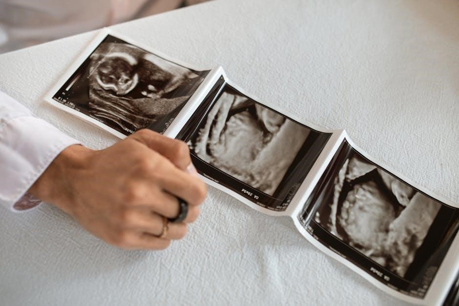
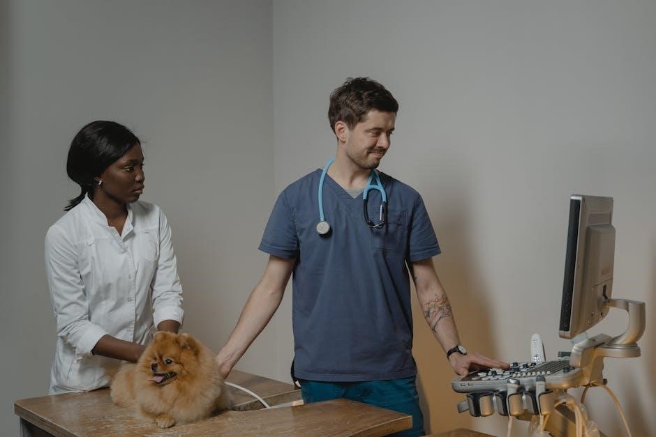
Ultrasound-guided cystocentesis is a minimally invasive procedure using real-time imaging to collect urine directly from the bladder. It ensures accuracy, minimizes contamination, and enhances diagnostic reliability in veterinary medicine.
Definition and Purpose
Ultrasound-guided cystocentesis is a minimally invasive veterinary procedure where a sterile needle, guided by real-time ultrasound imaging, is inserted into the urinary bladder to collect urine. This technique ensures precise needle placement, reducing the risk of complications; Its primary purpose is to obtain a sterile urine sample for urinalysis, which aids in diagnosing urinary tract infections, kidney disease, and other conditions. Additionally, it can relieve urinary retention caused by obstructions. The use of ultrasound enhances accuracy, especially in patients with non-palpable bladders or anatomical challenges, making it a valuable diagnostic and therapeutic tool in veterinary medicine. It is considered the gold standard for urine collection due to its safety and reliability.
Importance in Veterinary Medicine
Ultrasound-guided cystocentesis plays a pivotal role in veterinary medicine by enabling precise and sterile urine collection for diagnostic purposes. It is particularly valuable for patients with non-palpable bladders, such as obese animals or those with anatomical abnormalities. This method reduces contamination risks compared to catheterization or free-catch samples, ensuring more accurate urinalysis results. Additionally, it minimizes complications like urinary tract trauma, making it safer for pets with bleeding disorders or other health issues. The procedure is also therapeutic, aiding in relieving urinary retention caused by obstructions. Its reliability and safety have made it a cornerstone in veterinary diagnostics, especially for identifying conditions like urinary tract infections, kidney disease, and bladder abnormalities. Its versatility and efficacy make it indispensable in clinical practice.

How Ultrasound-Guided Cystocentesis Works
Ultrasound-guided cystocentesis uses real-time imaging to precisely guide a needle into the bladder, ensuring accurate urine collection by visualizing the target and surrounding structures.
Role of Real-Time Imaging
Real-time imaging in ultrasound-guided cystocentesis provides live visualization of the bladder and surrounding tissues. This allows precise identification of the bladder’s location, size, and position, even in challenging cases such as small or deep-seated bladders. The ultrasound probe generates high-resolution images, enabling veterinarians to select an optimal needle entry point, avoiding nearby organs or structures. This real-time feedback enhances accuracy, reducing the risk of complications like urinary tract trauma or contamination. Additionally, it is particularly beneficial for obese animals or those with irregular bladder shapes, where blind techniques may fail. Real-time imaging ensures safe and effective needle guidance, making the procedure more reliable and minimizing potential errors.
Guiding Needle Placement for Accuracy
Ultrasound-guided cystocentesis employs real-time imaging to direct needle placement precisely into the bladder. The ultrasound probe visualizes the needle’s trajectory, ensuring accurate penetration through the abdominal wall and into the bladder lumen. This guidance minimizes the risk of hitting surrounding organs or structures, such as the urethra or intestines. The ability to visualize the needle in real-time enhances control, particularly in challenging cases like deep-chested breeds or obese animals. Proper alignment and depth are maintained, reducing complications like urinary tract trauma or leakage. This precise placement also ensures the collection of a sterile urine sample, which is critical for accurate urinalysis results. The use of ultrasound significantly improves the safety and reliability of the procedure compared to blind techniques.
Preparation for the Procedure
Preparation involves patient positioning, sterile skin preparation, and ultrasound equipment setup. The bladder is located via ultrasound, ensuring proper needle placement and minimizing contamination risks during cystocentesis.
Patient Preparation and Positioning
Patient preparation involves positioning the animal to facilitate easy access to the bladder. Lateral or dorsal recumbency is commonly used, depending on the patient’s size and comfort. For obese animals or those with deep chests, lateral recumbency may be preferable. The area over the bladder is shaved and aseptically prepared to minimize contamination risk. Restraint is crucial, especially in fearful or uncooperative patients, to ensure accurate needle placement. Ultrasound imaging is then used to locate the bladder and guide the procedure. Proper positioning and preparation are essential for the safety and success of ultrasound-guided cystocentesis, ensuring a sterile and precise sample collection process.
Sterile Technique and Equipment Setup
Sterile technique is critical to prevent contamination during ultrasound-guided cystocentesis. The procedure requires aseptic preparation of the skin over the bladder with antiseptic solutions. Surgical gloves are worn to maintain sterility, and the ultrasound probe is covered with a sterile sheath. Essential equipment includes a 22-23 gauge needle (1-1.5 inches long) and sterile collection tubes or syringes. Ultrasound gel is applied to the probe to ensure clear imaging. The setup must be organized to allow seamless transition from imaging to needle insertion. Proper sterilization and organization of equipment minimize complications and ensure accurate, uncontaminated urine sample collection for urinalysis. Attention to detail in equipment preparation is vital for the success of the procedure.

Step-by-Step Process of Ultrasound-Guided Cystocentesis
The procedure involves locating the bladder via ultrasound, inserting a sterile needle under real-time guidance, and collecting urine for analysis, ensuring precision and minimizing complications.
Locating the Bladder Using Ultrasound
Locating the bladder using ultrasound is the first critical step in cystocentesis; The veterinarian uses a transducer to visualize the bladder in real-time, ensuring accurate identification. This method is particularly useful when the bladder is small, not easily palpable, or obscured by other structures. The ultrasound probe is moved over the abdominal area to identify the bladder’s position, avoiding nearby organs like the uterus or prostate. Clear visualization helps determine the optimal needle entry point, reducing the risk of complications. This step is essential for guiding the needle accurately and safely, especially in challenging cases such as obese animals or those with irregular bladder shapes.
Inserting the Needle and Collecting Urine
Under real-time ultrasound guidance, a sterile needle is carefully inserted into the bladder. The needle’s placement is monitored to ensure accuracy and avoid surrounding structures. Once the needle is correctly positioned, urine is gently aspirated into a sterile collection container. The procedure is typically quick, lasting only a few minutes, and minimizes contamination risks. Needle sizes, usually between 22-23 gauge and 1 to 1.5 inches in length, are selected based on the patient’s size. This method ensures a safe and precise collection process, reducing the likelihood of complications and providing a reliable sample for urinalysis.

Comparison with Blind Cystocentesis
Ultrasound-guided cystocentesis offers improved accuracy and safety compared to blind methods, minimizing contamination and complications. Blind cystocentesis is simpler but carries higher risks of contamination and inaccuracies.
Advantages of Ultrasound Guidance
Ultrasound guidance enhances the accuracy of cystocentesis by providing real-time visualization, reducing the risk of complications. It is particularly beneficial in patients with difficult anatomy, such as obesity or small bladders. This method minimizes contamination, ensuring sterile urine samples for urinalysis. Additionally, ultrasound guidance decreases the likelihood of puncturing surrounding structures, improving safety. It is especially useful in cases where the bladder is not palpable or when there are concerns about intra-abdominal diseases. Overall, ultrasound guidance makes the procedure more reliable and reduces the need for repeat sampling, making it a preferred technique in modern veterinary practice for optimal diagnostic outcomes.
Limitations of Blind Techniques
Blind cystocentesis relies on palpation and anatomical landmarks, which can be unreliable in patients with difficult anatomy. It poses risks like urinary tract hemorrhage, urine leakage, and bladder rupture. Obesity, small bladders, or intra-abdominal disease increase complications. Contamination risks rise without real-time imaging, potentially leading to inaccurate urinalysis results. Blind techniques lack the precision of ultrasound guidance, especially in challenging cases, and may result in incomplete or non-sterile samples. These limitations underscore the importance of ultrasound guidance for improving accuracy and safety in cystocentesis procedures, particularly in high-risk or complex patients.
Possible Complications and Contraindications
Complications include urinary tract hemorrhage, urine leakage, and rare bladder rupture. Contraindications involve bleeding disorders, pyometra, or bladder cancer, where cystocentesis may worsen conditions or spread disease.
Common Complications
Common complications of ultrasound-guided cystocentesis include urinary tract hemorrhage, which may contaminate the urine sample, and urine leakage into the peritoneal cavity, typically insignificant. Rarely, bladder rupture can occur, especially in animals with pre-existing conditions like urinary outflow obstruction. Hemorrhage is more likely in patients with bleeding disorders or those on anticoagulant therapy. Leakage usually resolves without intervention but may require monitoring. Bladder rupture, though uncommon, is a serious complication that necessitates immediate veterinary attention. Proper technique and patient selection are critical to minimizing these risks and ensuring the procedure’s safety and effectiveness.
Contraindications for the Procedure
Contraindications for ultrasound-guided cystocentesis include insufficient urine volume in the bladder, making the procedure ineffective. Patients with marked bleeding disorders or those on anticoagulant therapy are at higher risk of hemorrhage. Conditions such as pyometra, urinary bladder cancer, or transitional cell carcinoma pose risks, as the procedure may spread tumor cells. Additionally, animals with resistance to restraint or those unable to remain still during the procedure are not ideal candidates. Veterinarians must weigh these factors against the need for a sterile urine sample to determine if the benefits outweigh the potential risks.
Patient Selection Criteria
Patient selection for ultrasound-guided cystocentesis includes animals requiring sterile urine samples, those with palpable bladders, or cases where real-time imaging enhances accuracy and safety.
Indications for Ultrasound-Guided Cystocentesis
Ultrasound-guided cystocentesis is indicated for obtaining sterile urine samples for urinalysis, particularly in patients with non-palpable bladders, obesity, or deep-chested breeds. It is ideal for animals requiring accurate diagnostic sampling, such as those with urinary tract infections or kidney disease. Additionally, it is beneficial for patients with urinary outflow obstruction, as it helps relieve bladder distension. The procedure is also preferred in cases where blind cystocentesis may pose risks, such as in small or irregularly shaped bladders. Real-time imaging ensures precise needle placement, reducing complications and improving sample quality. This method is particularly advantageous in minimizing contamination and ensuring a safe, efficient procedure.
Patients Who Benefit Most from the Procedure
Patients with non-palpable bladders, such as obese animals or those with deep-chested anatomy, significantly benefit from ultrasound-guided cystocentesis. This procedure is particularly advantageous for animals with small or irregularly shaped bladders, where blind techniques may fail or pose risks. Additionally, patients with urinary outflow obstruction or those requiring precise diagnostic sampling, such as in cases of suspected urinary tract infections or chronic kidney disease, benefit greatly. Real-time imaging ensures accurate needle placement, minimizing complications and contamination. It is also ideal for patients where blind cystocentesis is challenging or unsafe, providing a reliable and sterile method for urine collection. This approach enhances diagnostic accuracy and patient safety in diverse clinical scenarios.

Equipment Required
Essential equipment includes an ultrasound machine with appropriate probes, 22-23 gauge needles, sterile collection materials, and ultrasound gel for clear imaging, ensuring accurate and safe procedures.
Ultrasound Machine and Probes
An ultrasound machine with high-resolution imaging capabilities is essential for guiding cystocentesis. Linear or curved probes, typically operating at 7.5–10 MHz, provide clear visualization of the bladder and surrounding structures. Portability and ease of use make modern machines ideal for clinical settings. The probe’s frequency ensures adequate tissue penetration while maintaining image quality. Real-time imaging allows precise needle placement, reducing complications; Recent advancements, such as compact devices like the SpeqView TM, offer cost-effective solutions without compromising image quality. Proper probe selection and machine setup are critical for accurate procedure outcomes, ensuring safe and effective urine sample collection in veterinary patients.
Needles and Collection Materials
The procedure requires sterile 22-23 gauge needles, typically 1-1.5 inches long, ensuring minimal discomfort and effective urine collection. Needle selection varies based on patient size, with longer needles for larger animals. A syringe or collection tube is attached to the needle, and sterile gloves are recommended to maintain asepsis. The equipment setup ensures a closed system, reducing contamination risks. Proper material preparation is crucial for obtaining accurate urinalysis results. The use of sterile supplies minimizes the risk of infection and ensures sample integrity. These materials are essential for the safe and successful completion of ultrasound-guided cystocentesis, providing reliable diagnostic outcomes for veterinary patients.

Advantages of Ultrasound-Guided Cystocentesis
Ultrasound-guided cystocentesis improves accuracy, reduces contamination risks, and enhances safety, especially in challenging cases like small or deep bladders. It minimizes complications and ensures reliable diagnostic results.
Improved Accuracy and Safety
Ultrasound-guided cystocentesis significantly enhances accuracy by providing real-time visualization of the bladder and surrounding structures. This reduces the risk of needle misplacement and potential damage to nearby organs or blood vessels. The use of imaging ensures precise needle placement, even in challenging cases such as small, deep, or irregularly shaped bladders. Additionally, it minimizes complications like urinary tract hemorrhage or bladder rupture. The procedure is particularly beneficial for obese animals, deep-chested breeds, or patients with abdominal disease, where traditional palpation may fail. By improving safety and reducing procedural risks, ultrasound guidance enhances the reliability of urine sample collection for diagnostic purposes.
Minimizing Contamination
Ultrasound-guided cystocentesis minimizes contamination by ensuring a sterile needle insertion directly into the bladder. Real-time imaging allows precise placement, reducing the risk of urine sample contamination from skin or other sources. This method is particularly advantageous compared to catheterization or free-catch samples, which are more prone to bacterial contamination. The procedure’s closed system and direct access maintain sample integrity, crucial for accurate urinalysis results. Furthermore, visualization helps avoid accidental aspiration of adjacent tissues or fluids, ensuring a pure urine sample. This reduces the likelihood of false positives in diagnostic testing and provides reliable data for clinical decision-making in veterinary medicine.

Clinical Applications
Ultrasound-guided cystocentesis aids in diagnostic urinalysis and therapeutic interventions. It supports accurate diagnosis and treatment, ensuring reliable sample collection for clinical decision-making in veterinary care.
Diagnostic Urinalysis
Ultrasound-guided cystocentesis is a critical tool for obtaining urine samples for diagnostic urinalysis. By directly accessing the bladder under real-time imaging, it ensures a sterile and accurate collection, minimizing contamination. This method is particularly valuable for identifying urinary tract infections, kidney disease, and other conditions requiring precise urinalysis results. The procedure allows veterinarians to gather samples from animals with difficult-to-palpate bladders, such as obese or deep-chested patients, enhancing diagnostic reliability. The use of ultrasound guidance also reduces the risk of sample contamination, providing clearer insights into a patient’s urinary health and aiding in timely, effective treatment plans. This approach is essential for achieving reliable diagnostic outcomes in veterinary medicine.
Therapeutic Uses
Ultrasound-guided cystocentesis is not only diagnostic but also serves therapeutic purposes. It is commonly used to relieve urinary retention or distension in patients with urinary outflow obstruction. By safely draining the bladder under ultrasound guidance, it helps reduce pressure and prevent complications such as bladder rupture. This procedure is particularly beneficial for animals with urethral blockages or neurological disorders affecting urination. Additionally, it can be used to decompress the bladder in critically ill patients, improving comfort and supporting urinary tract health. The precision of ultrasound guidance minimizes risks, making it an effective tool for both diagnostic and therapeutic interventions in veterinary care.

Future Trends in Ultrasound-Guided Cystocentesis
Advancements in ultrasound technology, such as portable devices and AI algorithms, will enhance precision and accessibility, making the procedure faster and more effective in veterinary practice.
Advancements in Ultrasound Technology
Recent advancements in ultrasound technology, such as high-resolution imaging and portable devices, are revolutionizing ultrasound-guided cystocentesis. These innovations improve visualization of the bladder and surrounding structures, enhancing procedural accuracy. Portable ultrasound devices, like the SpeqView™, offer cost-effective and accessible solutions for veterinary clinics. Additionally, the integration of artificial intelligence (AI) in ultrasound systems can aid in real-time guidance, reducing human error and improving safety. These technological improvements are expected to make ultrasound-guided cystocentesis more efficient and widely adopted in veterinary practice, ensuring better diagnostic outcomes for patients.
Emerging Techniques and Innovations
Emerging techniques in ultrasound-guided cystocentesis include the integration of artificial intelligence (AI) for enhanced imaging and real-time guidance. AI algorithms can improve needle placement accuracy and reduce complications. Additionally, advancements in probe design and ultrasound gel technology are enhancing image clarity and patient comfort. Portable ultrasound devices, such as the SpeqView™, are making the procedure more accessible in clinical settings. Innovations in needle design, including smaller gauges and echogenic tips, are also being explored to improve visibility and minimize discomfort. These advancements are expected to further streamline the process, making ultrasound-guided cystocentesis more efficient and reliable for veterinarians. Such innovations highlight the evolving nature of this diagnostic tool.
